Thursday, July 10, 2014

Cracked Tooth Syndrome (CTS)

What is CTS?
Minuscule cracks can form on your teeth, threatening your dental health. Why? 
  • Age plays a factor - as we get older, our teeth tend to weaken, making them susceptible to tiny hairline fractures that are not visible to the naked eye. 
  • We increase our chances of developing cracked teeth by exposing them to trauma, such as bruxism and chewing on hard objects.
  • When teeth have been heavily filled, it is not unusual that they develop cracked (or split) tooth syndrome (CTS). 
Symptoms include a sharp pain when you bite down into something hard. When you open your mouth and the teeth are no longer touching, the pain goes away. The pain does not usually linger after the biting action is finished. The fact that you feel pain when pressure is applied to the tooth means that the nerve is being affected. If the problem is not solved quickly, the nerve may die and the tooth will then require endodontic treatment (root canal).

Why does it hurt when I eat?
Pressing an object against the tooth opens the crack, causing the underlying dentin to move and irritating the pulp chamber (which contains the tooth's nerves). When the pressure is released, the crack immediately closes, causing a sharp pain. If left untreated, the pulp will eventually become damaged, and you will start to feel sensitivity to hot and cold or prolonged pain.

Common sites of cracked teeth
  • Premolars and molars, the back teeth that grind and crush food 
  • A tooth that has recently been drilled and a great amount of tooth structure has been lost
  • A tooth that has an extensive filling (usually silver amalgam) that has been in place for a long time 
  • Or a tooth that only has a small silver filling  
    • The filling has weakened the tooth just enough so that when you chew or bite, the tooth and filling separate slightly (flex), causing immediate and sometimes severe pain.
Types of cracked teeth:
  • Fractured Cusp: When the cusp (the raised section of the biting surface of your tooth) becomes fractured. If a fractured cusp does not break off on its own, it will need to be removed by a dentist and replaced by a dental crown.
  • Cracked Tooth: Run vertically, originating from the top part of the crown and working their way down. Treatment typically entails a root canal followed by a dental crown. If the crack has extended below the gum line, the tooth may require a tooth extraction. 
  • Split Tooth: When a cracked tooth is not treated, the crack can extend beyond the root, causing the tooth to split. Although these teeth are difficult to save, they can sometimes be treated with a root canal. 
  • Vertical Root Fracture: Sometimes the crack starts at the bottom of the root and works its way up. If caught early, endodontic dental surgery may correct the situation. 
Treatment
Vertical Fracture

  • Treatment will involve at least one x-ray to assist in the diagnosis and to rule out other causes. We will try to find the section of the tooth that is causing the problem by pushing on the various sections of the tooth or having you bite on a hard object.  
  • When the section of the tooth that is cracked is found, it makes treatment easier. First, the tooth is anesthetized and the old filling is removed. 
  • Then we carefully inspect the area to determine whether the cracked section can be seen. Very often it is visible at this point. 
  • The next step is to see whether the split area can be fixed with a direct filling (bonded). This is the ideal situation if the crack is small.  If the crack is small enough, it may be removed by replacing the filling. Bonded white fillings and bonded amalgam filling will hold the tooth together making it less likely to crack
    • Crown
    • Your dentist may first place an orthodontic band around the tooth to keep it together. If the pain settles, the band is replaced with a filling that covers the fractured portion of tooth (or the whole biting surface).
  • Unfortunately, this rarely occurs. If the crack goes too far vertically, there is a possibility the tooth may need to be removed and replaced with an artificial one (bridge, denture, or implant). More often (over 95% of the time), the biting surfaces of the tooth must be entirely covered and protected first with a provisional (temporary) onlay or crown. 
  • If this is successful in eliminating the pain (we usually wait for a few weeks to be sure the problem is resolved), an impression for a laboratory-fabricated casting - either a porcelain or resin onlay or a crown - is made. If adequate tooth structure remains, a partial coverage restoration - an onlay - is preferred. If the tooth has been badly cracked or if not much tooth remains, then a crown will be necessary. The purpose of either type of cast restoration is to unite all sections of the tooth so it cannot move or separate under normal biting forces. If the provisional restoration is successful in eliminating the pain, we expect that the final cast crown or onlay will correct the problem. 
  • The nerve may sometimes be affected so badly that it dies. Root canal treatment will be required if the tooth is to be saved.
The earlier a split tooth is diagnosed and treated, the better success there is in treating it. If the split is severe, a root canal may be necessary to save the tooth. Depending on the symptoms you describe, we may choose to treat it as a bite adjustment, small filling, or larger preparation for an onlay or  crown. If these do not work, the treatment plan will be modified. If left untreated, the tooth may eventually be lost. It is also possible that the initial split of the tooth may be such that it cannot be saved, despite our best efforts.

Delaying Treatment
What happens if CTS is not treated quickly? The best you can hope for is that the tooth continues to hurt only when you chew or bite. This does not often happen. Usually, the broken section of tooth gets weaker and weaker until it fractures off. Additionally, if the crack gets deeper into the tooth, the nerve will die and the tooth will need endodontic treatment before the crown or onlay is placed. Sometimes the nerve is immediately affected by the initial split and dies. This may occur quickly or may take years before it is evident. Every case of CTS is unique. If it is any consolation, the cracked tooth is not your fault. It is a result of your teeth being drilled and filled with big silver fillings when you were younger. We see this particular dental problem mostly in patients who are between 25 and 45 years old.

Unfortunately, cracked teeth do not go away. Many times, they only hurt when you bite on them from one particular angle. If the fractured segment is not stressed, the tooth feels normal. You might also be able to "train" yourself to chew on different teeth and avoid the cracked tooth. At best, you only postpone necessary treatment while the nerve may be slowly dying.

After Treatment
If treated by adjusting your occlusion (bite) on the tooth
This will lessen the symptoms. If the discomfort you feel when you bite is not eliminated within one week of treatment, please call our office and let us know. If the split is still present, a different approach will have to be tried. Give the tooth a few days to "calm down" before you try to bite down on hard foods.

If treated by removing the old restoration and placing a bonded filling in its place 
If the split is small, this may eliminate the problem. Do not bite down hard on the tooth for a few days, then gradually place more pressure on the tooth. Call our office in one week and let us know how it feels. If the split is still causing a problem, a different approach must be tried. If left untreated, the tooth may split further and you may need endodontic treatment and a crown. If the tooth splits severely, it may have to be extracted.

If treated by cementing a temporary crown onto the cracked tooth with temporary cement 
Because the crack was so severe, this procedure was used to determine whether or not the tooth can be treated without performing a root canal. Please give the tooth several days of rest before you try biting down on hard foods. Expect the tooth to be sensitive for a few days after the temporary is placed. This is normal. Gradually apply more force when you bite into foods, and gradually try to eat harder foods. Expect the temporary crown to remain in place for several weeks, until it can be determined whether or not the problem is solved.

If you have any questions about cracked tooth syndrome, please feel free to ask us.

Wednesday, July 9, 2014

Foods That Cause Tooth Decay

Cariogenic: causing tooth decay.
Foods (such as candy and sweets) that contain sugar and other carbohydrates that can be metabolized by bacteria in plaque are described as cariogenic

How long food remains in mouth:
  • Sugary liquids, such as soft drinks, leave the mouth quickly and are not as cariogenic
  • Sticky foods, such as raisins or caramels, adhere to the teeth and stay in the mouth longer and are more cariogenic
Frequency vs. Amount:
  • Someone who snacks all day on small amounts of cariogenic food is far more likely to develop decay
  • Someone who eats a larger amount of cariogenic food, but only once a day, is less likely to develop decay 
Refined carbohydrates vs. Complex carbohydrates:
  • Foods such as crackers, although not sweet, are cariogenic because they contain refined carbohydrates that stick to the teeth. They remain in the mouth long enough to be broken down into sugars that can be used by the bacteria in plaque.
  • Complex carbohydrates, such as fruits and vegetables, are less cariogenic because they clear the mouth before they are converted into simple sugars that can be used by bacteria.  
Flow of saliva to speed clearance of food from mouth and provide a source of dietary fluoride to strengthen the tooth and assist in remineralization
  • Sugarless gum or hard candy, as well as sipping water frequently, will increase the saliva flow
  • Various medicines can cause a dry mouth, and try to avoid spicy or salty foods

Tuesday, July 8, 2014

Give Yourself A 60-Second Dental Check-Up

Regular visits to your dentists are absolutely essential to prevent serious dental problems. But it is also important to give yourself an oral check-up from time to time. This simple process just takes a minute, but it can yield valuable clues to the health of your mouth, teeth, and gums.

Stand before a well-lit mirror and open your mouth wide.
1. Check your gum line and the lines between your teeth
Good: There should be no brown lines here
Bad: If you see brown lines, that is a sign of tartar build-up
What to do: Time to go to the dentist. Once the brown lines are cleaned up at the clinic, regular flossing will keep tartar away.

2. Stick out your tongue. What does the surface look like? 
Good: A healthy tongue should be pink
Bad: If it is coated with white, it can be an indication of poor digestion. This coating also results in bad breath.
What to do: Use a stainless steel scraper to clean your tongue every morning. 

3. Are there ridges along your tongue? 
Bad: This is often a sign of tooth grinding. People may not be aware they grind their teeth, but it can happen when they are stressed. 
What to do: If you wake up with an aching jaw, you should see a dentist.

4. Take a look at the inside of your cheeks. 
Bad: Do you see white patches? These can occur if you chew the inside of your mouth when you bite and chew your food. 
What to do: These should be checked by your dentist because a wound inside the cheek can not only get infected, but may also be a sign of pre-cancerous changes, especially in smokers. An ulcer that does not heal within three weeks should also be investigated.

5. How do your gums look?
Good: If they are pink and rosy, your gums are in good health. 
Bad: But if they are red, it is a signal that gingivitis or gum inflammation has turned into gum disease. Another clue: gums that bleed when you brush your teeth. 
What to do: Your dentist can help you with a course of antibiotics and dental hygiene guidance on how to rebuild gum health. 

Bad: Pale gums can indicate iron deficiency - a warning sign of anemia. 
What to do: Up your intake of green vegetables, tofu, chickpeas, lentils, and dried fruits such as raisins and prunes.

Monday, July 7, 2014

Am I Grinding My Teeth At Night?

How do you find out if you are grinding your teeth at night? Many patients are often unaware that they have this problem. Some people do grind or clench their teeth during the day, but it oftentimes occurs at night when the person is asleep. Because of this, it can be difficult to self-diagnose.

Ways to find out
  • When you wake up, are you in pain?
    • Such pressure applied can cause a tooth to hurt or cause soreness in the jaw muscles.
  • Does someone observe you grinding your teeth while you are sleeping?
    • Many people who grind their teeth at night will make repetitive clicking or grinding noises while sleeping. They will not make these noises all night long, but instead exhibit repeated short periods of grinding/clenching activity.
  • Does your front teeth look worn when you look in the mirror?
  • Do you experience these symptoms when you wake up?
    • A dull, constant headache
    • A sore jaw
    • Audible teeth grinding sounds as you are falling asleep
    • Tooth sensitivity to heat, cold, or brushing
    • Inflammation of the gums (gingivitis)
    • Wounds on the inside of the cheeks (from biting)
Ask your dentist because chronic teeth grinding can result in fracturing, loosening, or the loss of teeth.Your dentist can observe the wear patterns on your teeth and examine your mouth and jaw for signs of grinding, such as jaw tenderness, worn-down teeth, and other abnormalities. They will also check to make sure that you are not suffering from some other ailment that cause similar pain, such as: dental disorders, ear disorders, or TMD.

How to fix
  •  Ask your dentist
    • Dentists often recommend that patients wear a night guard in order to protect teeth and jaws from further damage. Many patients who do wear a night guard report that they feel better in the mornings. A previous blog post goes over the process of getting a bite guard.
    • Muscle relaxants can be prescribed
    • Crowns or onlays can be put onto your teeth to fix any damage caused to the teeth and repair your bite
  • Understand bruxism: it is a condition where a person grinds, gnashes, or clenches his teeth unconsciously. 
  • Reduce your stress. The degree of severity can be influenced by the amount of stress that someone is experiencing. Many patients who are aware of their nocturnal bruxing report that it can be worse during stressful periods of their lives.
    • Stress counseling, exercising, meditating
    • Eliminate major sources of stress in your life
    • Go to bed and wake up at the same time every day
    • Have fun with family and friends
    • Eat well: three well-balanced and healthy meals each day
    • Relax before bed
  • Reduce stress on your mouth
    • Stop chewing non-foods (chewing on pencils)
    • Train yourself not to clench your jaw during the day
  • If your muscles feel sore, you can apply ice to your jaw to alleviate the pain
  • If your jaw hurts, you can take a pain reliever like ibuprofen for temporary relief

Thursday, July 3, 2014

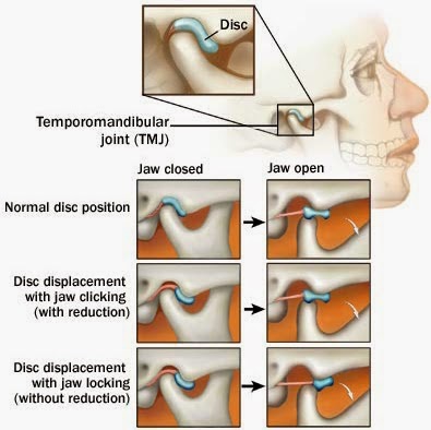
Does your jaw click when you open your mouth?

If you experience a clicking jaw when you open your mouth, or if your jaw locks, you may be experiencing problems with you temporomandibular joint. Problems with your jaw joint, known as the temporomandibular joint (TMJ) are considered a part of temporomandibular disorder (TMD).

No pain
If you do not experience any pain with it, you do not need an invasive procedure to correct it. You can try wearing a well-made night guard at night.

Pain
If you do experience pain associated with your clicking jaw, then the joint may be inflamed. Try to avoid over-working your jaw: eat easy-to-chew foods, do not open your mouth wide. Keep in mind that other conditions cause similar symptoms to TMD, such as a toothache, sinus problems, arthritis, or gum disease. See your dentist for a diagnosis and to receive treatment for this problem.

A previous blog post outlines the symptoms often associated with TMD 

Discomfort
Jaw surgery is usually not necessary. Occasional discomfort in the jaw joint and chewing muscles is quite common and usually not a cause for concern. If you are diagnosed with TMD, you can use simple self-care practices to relieve discomfort:
  • eat soft foods
  • do not open your mouth wide
  • do not chew gum
  • apply heat or ice packs
  • avoid extreme jaw movemetns
  • nonsteroidal anti-inflammatory drugs
  • muscle relaxants
  • physical therapy 
  • wear a night guard at night

If you have any questions about TMJ or your jaw joint, please feel free to ask us.


Wednesday, July 2, 2014

Classification of Periodontal Disease

Any periodontal disease is undesirable and, if left untreated or ignored, can lead to a number of serious dental problems. If you wish to maintain your teeth and gums (gingiva) in a healthy and disease-free state, it is important that you brush properly and use dental floss daily. Do these procedures as we have instructed. Return for continuing dental hygiene care at the time intervals that we have recommended. These time intervals for your cleaning appointments have been established specifically for your existing dental condition. The intervals can and will fluctuate according to your ability to take care of your teeth and gums. A periodontal infection is site-specific and episodic in its nature. Any delay in your office-related routine dental hygiene recare appointments could prove detrimental to your oral health. 

What is periodontal disease? Periodontal disease is an infectious process classified according to how much damage has been done to the structures surrounding the teeth, namely the gingiva (gums) and bone. It is an infection in your mouth. The better you clean your teeth to remove all the plaque bacteria, the less likely you will be to develop periodontal disease. A previous blog post details how the disease progresses and how to solve it.

The following is a brief overview of the American Academy of Periodontology's classification of the types of periodontal disease.

Type 1 - Gingival Disease: An inflammation or lesion of the gum characterized by changes of color, gingival form, position, surface appearance, and presence of bleeding and/or pus.


Type 2 - Chronic Periodontitis: An inflammation of the supporting structures of the teeth associated with plaque and calculus; the rate of progression is affected by local, systemic, or environmental factors. It can be further classified as localized or generalized.


Type 3 - Aggressive Periodontitis: Characterized by a rapid rate of periodontal disease progression in an otherwise healthy individual in the absence of large accumulations of plaque and/or calculus. It can be further classified as localized or generalized.


Type 4 - Periodontitis as a Manifestation of Systemic Disease: Periodontitis associated with blood or genetic disorders.


Type 5 - Necrotizing Periodontal Disease: Ulcerated and necrotic gums between the teeth and at the tooth margins. It can be further classified as necrotizing ulcerative gingivitis or necrotizing ulcerative periodontitis.


Type 6 - Abscesses of the Periodontium: A localized pus-forming infection of the periodontal tissue.


Type 7 - Periodontitis Associated with Endodontic Lesions: Localized deep periodontal pocket extending to the tip of the root of the tooth involving pulp death.

Type 8 - Developmental or Acquired Deformities and Conditions: Gingival disease or periodontitis started by localized tooth-related factors that modify or predispose to plaque accumulation or prevention of effective oral hygiene measures.

Due to the nature of the disease, most classifications will involve both a generalized and a localized diagnosis. 

See this previous blog post for early detection signs of periodontal disease.

If you have any questions about the classification of your periodontal disease, please feel free to ask us.

Tuesday, July 1, 2014

Maryland Bridge (Bonded Resin Retainer)

The Maryland bridge, or bonded resin retainer, is an alternative to implants and conventional crowns to replace missing teeth. This type of replacement procedure can be considered when the space to be restored is next to teeth in good alignment that are not heavily restored with filling materials. Because this type of bridge is adhesively bonded to the enamel on the abutment teeth, having adequate enamel is important. A Maryland bridge is fixed into place and is not meant to be removed at night.

Maryland Bridge vs. Fixed Bridge
One advantage of this process over a conventional bridge is that a missing tooth can usually be replaced with a minimum amount of preparation (drilling) of the remaining teeth. It is not necessary to prepare the tooth to the same extreme degree as with full tooth coverage restorations (crowns or caps). This method is an extremely conservative procedure. It is also less expensive (about one quarter less) than a conventional bridge. Tissue health around the abutment teeth is usually excellent because little or no preparation of the tooth structure near the gingival (gum) tissues is needed. Unlike conventional full coverage restorations, teeth prepared for a Maryland bridge retainer will not need endodontic treatment (root canal therapy) at a later date.

If the patient has abusive eating or bruxing (grinding) habits, this type of restoration is contraindicated. Although we do consider it more of a restoration for posterior (back) teeth rather than anterior teeth, it does display metal more so than a conventional bridge. The biggest disadvantage of a Maryland bridge is that the expected life may not be as long as that of a conventional bridge made of full coverage crowns. This technique was only introduced in the early 1980s and therefore does not have the extensive successful history of full coverage bridges. While breaking of a Maryland bridge is not likely, debonding can occur, requiring a re-cementation. In some rare instances, it might have to be redone. In either case, you may be charged a fee for the procedure. A Maryland bridge is a fixed replacement for a missing tooth, but one that will need to be redone in the future. The success rate at 5 years of Maryland bridges in our practice is approximately 80%. However, this is a much better result than the national average, which shows a success rate of about 33% at 4 years. Maryland bridges are most appropriate for single-tooth replacement. 

If dental implants are not an option for you for tooth replacement and you do not want the teeth on either side of the space to be radically prepared (cut down or drilled), then you should consider the Maryland bridge technique. For many patients, the advantages of less drilling of the teeth and the reduced cost make it a desirable alternative.

If you have any questions about the Maryland bridge technique, please feel free to ask us.