Showing posts with label periodontal disease. Show all posts

Friday, January 27, 2017

Periodontal Disease

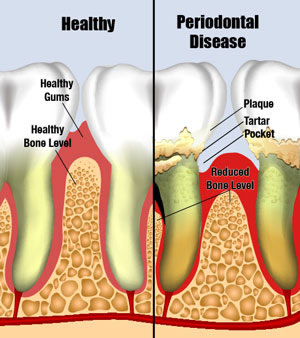
Periodontal disease is an infectious process classified according to how much damage has been done to the structures surrounding the teeth, namely the gingiva (gums) and bone. It is an infection in your mouth. It can happen anytime, around your teeth, affecting some or many of your teeth to varying degrees. There are genetic predisposing factors to periodontal disease, and our immune systems play a role in gum health, but it is usually related to how well you are able to keep your teeth clean through proper oral self-care. The better you clean your teeth to remove all the plaque bacteria, the less likely you will be to develop periodontal disease.

Progress of the Disease
The bacteria that cause this disease first cause the gum tissue to become inflamed and pull away from the teeth. As the problem becomes more serious, the bone that supports the teeth also becomes infected and begins to break down and dissolve. The teeth then become loose. Once the bone disappears, it is extremely hard, if not impossible, for new bone to be rebuilt. The damage is permanent and your teeth, the surrounding bone, and your general health will be compromised.
Periodontal disease is classified into several types. The mildest form of this infection will show up in red and swollen gum tissue that bleeds easily. There is seldom any pain involved at this stage. You may also notice that your breath becomes offensive and you feel the need to use mouthwash. Our sense of smell does become immune to the same odor, so we can lose our ability to detect our own offensive, diseased breath. As the disease progresses, the gum tissue becomes more red and swollen, more bleeding can be seen, and the teeth begin to become loose. This tooth mobility is a sign that there is a severe problem. There may still be no pain at this advanced stage. As more and more bone is lost and more teeth become involved in the infection, it becomes harder to treat. At this point, many times, the management of your problem will involve periodontal surgical procedures. If this is the case, you may be referred to a periodontist (gum specialist) for further treatment. Most of the time, periodontal disease starts and continues because of neglect. Brushing and flossing of teeth are not being done effectively on a daily basis. You may have been neglectful in getting your teeth checked and cleaned within the time frame intervals you need. Once we have diagnosed the disease, we will inform you of the problem and suggest treatment. If treatment is not completed, however, the disease will continue to progress. Unfortunately, the disease is quite invisible to most people until severe and possible irreversible damage has occurred.

Solution
If it has been diagnosed in the early stages and has not progressed to bone loss, a proper cleaning (prophylaxis) will solve the problem. Scaling and root planing over multiple appointments may be needed for more advanced cases. In the most advanced cases, periodontal surgery and tooth loss are inevitable. You will receive an estimate of fees for the recommended treatment.
Periodontal disease is a condition that must be treated quickly. We believe that if the infection is aggressively treated in its early stages, conservative periodontal treatment may be possible and effective. Although we do not automatically rule out periodontal surgical intervention, we hope you can either avoid it or reduce the amount you will need.

Successful treatment of your periodontal problem will depend on several factors. But the most important of these is your ability to perform excellent oral self-care—brushing, flossing, and the use of periodontal aids—on a routine, daily basis. Without this, periodontal treatment will fail, and the disease will return. 

Tuesday, August 12, 2014

Scaling and Root Planing

What is scaling?
Scaling is a periodontal dental procedure in which plaque and calculus are removed from the tooth both above (supragingival) and below (subgingival) the gum (gingiva)

What is root planing?
Root planing is a procedure in which diseased or altered portions of the root surface, the cementum, and dentin are removed and the resulting new surface is made smooth and clean. The more altered and damaged the root surface has been from calculus (tartar) accumulation, the more the need for root planing.

Why is it necessary?
The purpose of scaling and root planing is to remove all debris from the teeth. Any item that can cause inflammation of the gum tissue must be eliminated. The root surface must be made as smooth as possible. Irregularities in the root surface can contribute to gum inflammation. Irregularities are sites for bacteria and plaque buildup. The bacteria and the toxins they produce in the plaque are held against the tooth by the calculus. In this way, plaque and calculus on the teeth have been positively linked to gum disease.

What is the procedure like?
Depending on the severity of your particular periodontal problem, scaling and root planing may be the definitive treatment and no further procedures will be required. In many cases scaling and root planing are only a necessary part of the overall therapy. It is a demanding procedure. It requires much more time than the familiar adult prophylaxis (cleaning). It is usually done in multiple appointments, treating a quarter, half mouth, or your full mouth at each appointment. In this office we find that most patients are most comfortable if the area to be treated during the root planing procedure is anesthetized with a local anesthetic.

Recare
The scaling and root planing may have to be repeated in the future. It is customary to place the patient on a 3- to 4-month hygiene recare schedule. Scientific evidence clearly shows that for individuals who have demonstrated a predisposition to periodontal disease, an interval of 6 months is too long. We will determine the appropriate interval for you. As your situation changes, there may be changes in the length of these intervals as well.

What happens after treatment?
Other than the teeth being somewhat sensitive after the scaling and root planing procedure, there is little postoperative discomfort. The sensitivity will diminish with time. If you have been diagnosed as having severe periodontal infection, the sensitivity may remain for quite some time and further procedures may be needed to eliminate sensitivity. Although many procedures in dentistry can be considered elective, we consider scaling and root planing to be a necessity for your dental health.

Post-procedure Tips:
  • If therapeutic prophylaxis has been completed because your gingival (gum) tissue showed signs of infection and inflammation, and if you had significant calculus (tartar) buildup:
    • You may notice that your teeth feel different where the calculus was removed. The soft tissues may be sensitive or sore for approximately one day as they begin to heal. You may find that taking an over-the-counter pain reliever (aspirin, ibuprofen, etc.) will help during this 24-hour period. You may also rinse your mouth every few hours with warm salt water. Make sure that you brush and floss your teeth during this time period as you have been instructed. be gentle, because the brushed areas may be sore, but be thorough! You do not want to have the periodontal infection begin again.
  • If scaling and root planing, or other more involved periodontal procedures has been completed:
    • You can expected your gingival (gum) tissues to be quite sore. This is normal when the gum tissues have been infected and inflamed for some time. The more severely they have been affected, the more discomfort you can expect. This soreness should go away very quickly. You may rinse with warm salt water every few hours until the soreness is gone.
  • If scaling and root planing has been completed:
    • You may also notice that the teeth have become sensitive to temperature changes after the scaling and root planing. This sensation frequently occurs when the surfaces of the roots of your teeth have been cleaned. Removal of the debris covering the roots and attached to the roots leaves the roots open to temperature stimulus. If the problem persists, please let us know.
  • When you examine your gums closely in a mirror, you will also observe that the color, texture, and position of your periodontal tissues will undergo a change as the healing takes place. The swollen, reddened gum tissue will shrink, become more firm, and return to a healthy pink color. Watch for these welcome signs of improvement and be encouraged by the healing process.
  • Please do not forget to brush, floss, and use other periodontal cleaning aids as you have been taught. It is important that you begin establishing proper oral self-care habits immediately. If you find that the recently treated areas are sensitive to the brushing and flossing, be gentle - but be thorough! With proper technique you cannot damage the teeth or gingival tissues.
  • Brush after ever meal with a fluoride-containing toothpaste. Rinse with a fluoride-containing mouthrinse once each day.
Preventing Recurrence
Once scaling and root planing has been completed, it is most important for you to practice the brushing and flossing techniques in which you will be instructed. If we have recommended any additional periodontal aids, you must use them, too. Your cooperation is vital if the procedures are to be successful. To remain disease-free, you will need to remain constant in your oral self-care regimen.

If you have any questions about scaling and root planing, please feel free schedule a comprehensive dental exam with our office at 512-250-5012.

Tuesday, August 5, 2014

Splinting Teeth

Healthy Teeth Are Strong and Steady
In their normal state, teeth surrounded by healthy supporting structures exhibit very little mobility. Mobility can be defined in this case as movement of the teeth. Pushing on the teeth with dental instruments may cause the tooth to be deflected slightly from the "at rest" position, but this movement will be very, very slight.

Why Teeth May Need To Be Splinted
When the supporting bone is compromised and affected by periodontal disease, the teeth will show more mobility. If the tooth or teeth are subjected to trauma, they can be loosened in their sockets. Bruxing and grinding habits can also loosen teeth.

Teeth that are not too severely damaged by trauma will return to their former stability. Temporary splinting of the loosened teeth to each other or to other undamaged teeth may be required.

If the mobility is caused by clenching or grinding of the teeth, adjustment of the bite (occlusion) and the fabrication of a protective antigrinding/bruxing appliance may be indicated. In this case, no splinting of the teeth would be required.

The most common reason for splinting teeth is mobility cause by periodontal disease. The teeth show more movement as the bone support for the teeth diminishes. Multi-rooted teeth (molars) often show less mobility than single-rooted teeth with the same amount of bone loss. But the need for treatment is just as important. The more mobile the teeth, the more damage has been done, and the more splinting will be necessary.


The Splinting Procedure
Fixing periodontal disease
The first step in elimination of tooth mobility is to begin to correct the periodontal problem. If the teeth are mobile, the periodontal problem is probably advanced and the corrective measures could be both involved and time-consuming. Splinting may be started immediately. It involves attaching the mobile and perhaps non-mobile teeth together with a wire, acrylic, or a combination of the two. Attaching the teeth together gives them all more strength. Splinting has a limited life expectancy and must be repaired or replaced periodically. There is often a fee separate from the initial splinting fee associated with these procedures. You will be informed as to what your particular condition requires for short- and long-term therapy.

A more extensive form involves splinting the teeth together with cast and cemented restorations - crowns, bridges, bonded metal retainers, etc. This type of splinting will last much longer and is more expensive. The purpose is the same as that of external splinting - to attach the mobile teeth together so that they derive more strength and move less.

Teeth that are splinted will also require different and more involved brushing and flossing on your part. We will demonstrate these procedures for you.

Fees
Costs for splinting procedures vary greatly. It will depend on the number of teeth to be splinted, severity of the mobility, prognosis of the teeth, and the type of splinting selected.

If you have any questions about splinting teeth, please feel free to ask us.

Wednesday, July 30, 2014

Sealants and Fluoride: Benefit to Adult Patients

Dental decay can develop at any time, regardless of a person's age. A change in diet, change in lifestyle, change in oral self-care habits, the use of prescription medications, or a change in systemic health due to the normal aging process can all affect the caries (decay) susceptibility. Few people remain completely free of decay. Proper oral self-care on your part and properly spaced dental hygiene prevention appointments will go a long way to reduce the opportunity to have new decay to begin.

As you age, it is possible that some of your gum tissue will recede, exposing the root surfaces of your teeth. This gum recession can occur from improper brushing (brushing too hard with a hard toothbrush) or as a result of past periodontal problems. The more a tooth and root are exposed, the greater is the surface area you will have to keep clean. Sometimes the teeth with exposed roots are very hard to keep clean. These roots may be sensitive to temperature changes and are often times uncomfortable to brush. Decreased salivary flow (dry mouth) helps to create a breeding ground for bacteria to accumulate on the enamel and especially on the root surface. And root decay usually progresses quite quickly!

Goal of Prevention
Your goal should be to keep the dentist from drilling your teeth. Any reasonable preventive measure that is available should be seriously considered. When the dentist drills, you lose. When the dentist does not drill, you win.
                               
                                                         Dental Sealants
Please see our blog post on sealants. Although sealants are primarily designed for children, adults who have a history of active decay should consider having sealants placed on the posterior (back) teeth where indicated. We will tell you where it is possible to place the sealants. Even if you have not had a cavity for a long time, consider the application of a sealant as an inexpensive insurance policy for your teeth. Perhaps you would never get decay on the unsealed surfaces. But, just as you insure your home against destruction by fire, a sealant insures the tooth surface from decay. Preventive measures may allow you to avoid having your teeth drilled. You win! 

Topical Fluoride
For a similar reason, we advise the use of topical fluoride treatments for adults. The effectiveness of systemic and topical fluoride in preventing decay is well documented. When a cavity first starts, an application of fluoride might (depending on when it is used) reduce or eliminate the need for drilling.
Fluoride Varnish

An alternative to the fluoride treatment we can provide in our office is a daily rinse. If you can rinse with an over-the-counter mouth rinse containing fluoride every night as directed on the rinse label, you do not need the office topical fluoride treatment. If you cannot rinse daily as instructed, you will need the benefit from the strong office-applied topical fluoride treatment. Your oral health will benefit most from small increments of fluoride that are applied daily rather than one larger concentration every 6 months. However, only you know whether you will be faithful in your rinsing routine. When in doubt, let us do it here.

We stress prevention of dental disease above all else. Sealants and topical fluoride treatments are two of the more important preventive dental measures that we believe will significantly enhance your oral health.

Monday, July 14, 2014

Extractions

Reasons for Recommending Tooth Extraction
Teeth may need to be extracted for several reasons, including but not limited to:
  • severe periodontal disease
  • irreversible damage to the nerve tissue inside the tooth (and the patient decides against saving the tooth)
  • failed endodontic therapy
  • extreme fracture or decay of the tooth structure
  • improper positioning of the tooth or for orthodontic purposes
To a great extent, the reason for the extraction will influence the amount of discomfort you might experience subsequent to the procedure. When the tooth is to be extracted for periodontal reasons, there will be reduced bone support for the tooth and the tooth might be removed more easily than if there were full bone support. In this case, there might be lessened discomfort following the extraction.

The Procedure
1. Numb the tooth: Your dentist will need to anesthetize (numb up) both your tooth and the bone and gum tissue that surround it. This is done as an injection.

2. Extraction: The root portion of a tooth is firmly encased in bone (its socket), and tightly held in place by a ligament. During the extraction process, the dentist needs to both "expand the socket" (widen and enlarge it) and separate the tooth from its ligament, to the point where the tooth is loose and free to come out.
  • What does it mean to "expand" a tooth's socket? If you have ever tried to remove a tent stake that has been driven deeply into the ground, you know that you can't just pull the stake straight up. Instead, you first have to rock the stake back and forth, widening the hole in which it is lodged. Once the hole has been enlarged enough, the stake will come out easily.
  • How does the dentist expand the jaw bone? The bone inside the jaw is spongy. When a dentist applies firm pressure to a tooth (forcing it against the side of its socket), the bone will compress. After repeated application of pressure, from many different angles, the entire socket gradually becomes expanded. At some point, enough space will have been created (and the ligament separated from the tooth enough) that the tooth will come out.
3. Closing the extraction site: Once your tooth has been removed, your dentist will begin the process of closing your extraction site. This may include:
  • Removing infected or pathologic tissue by curetting (scraping) the walls of the tooth socket
  • Using finger pressure to re-compress the "expanded" socket
  • Rounding off sharp bone edges
  • Evaluating the tooth socket for sinus complications (upper back teeth)
  • Washing out ("irrigating") the socket, so to remove any loose bone or tooth fragments that remain
  • Placing stitches (usually only after surgical extractions).
  • Placing folded gauze over your extraction site and then having you bite down on it so to create firm pressure
The Tools
1. Dental Elevators: During the extraction process, a dentist will usually use an elevator first. These instruments are designed to be wedged in the ligament space between the tooth and its surrounding bone. As the elevator is forced and twisted, the tooth is pressed and rocked against the bone. This helps to expand the socket. It also helps to separate the tooth from its ligament. As this work is continued, the tooth will become more and more mobile. In some cases, the elevator may be able to shove the tooth on out. If not, the dentist will switch to the use of extraction forceps and remove the tooth with them.

2. Extraction Forceps: A dentist will usually keep a number of different extraction forceps on hand. Each one will have a design that's been specially made to grasp a certain type of tooth. When they're used, the dentist will grasp the tooth with the forceps and them firmly and deliberately rock it back and forth as much as it will. Because the bone that surrounds the tooth is compressible, the socket will expand. In addition to a rocking motion, a dentist will also rotate the tooth back and forth. This twisting action helps to rip and tear the tooth from the ligament that binds it in place. At some point, the socket will be enlarged enough, and the ligament torn enough, that the tooth can be easily removed.

What will I feel?
1. Pressure
2. You should not feel any pain 
3. Expect to hear startling extraction noises: These are often just routine

Following the Extraction
We will tell you the reason for the extraction and let you know what to expect following the procedure. Please follow the instructions given to you. If antibiotics are prescribed, take them until the prescription is completely finished. If pain medication is prescribed, take it only if necessary. If the medication prescribed contains a narcotic component, such as codeine, do not drive a motor vehicle or operate machinery that could prove dangerous to yourself or others. Expect some bleeding to occur from the extraction site for the first 24 hours. Remember, there is now a hole in your jaw from which the tooth has been removed, and the hole can be quite large. Some bleeding is to be expected.
  • Do not spit, rinse, or smoke for 24 hours
  • Do not drink through a straw for 24 hours
  • It would be a good idea not to brush near the extraction site for a day or two
  • When you brush and floss the area, be gentle!
  • For 24 hours after the extraction, try to chew food away from the extraction site
  • Some slight swelling in the area is to be expected, especially if the extraction was difficult
  • If sutures are placed, return to have them removed
  • If medication has been prescribed for possible post extraction discomfort, take it as directed
  • If prescription medication has NOT been given, you may take your usual over-the-counter pain reliever, as directed
 Please notify us if:
  • There is extended bleeding from the extraction site. Slight bleeding for several hours is normal.
  • Anything other than slight swelling occurs.
  • Discomfort continues for more than 24 hours, especially if it is not relieved by over-the-counter pain relievers.
Some infrequent complications of routine oral surgical procedures include (but are not limited to):
  • fracture of adjacent teeth or restorations (which of course would mean that these affected areas must be restored to normal function after the healing of the extraction site)
  • separated root tips or root fragments
  • temporary or permanent nerve damage to the area, resulting in anesthesia or paresthesia (numbness)
  • incomplete healing, resulting in severe pain - a "dry socket"
  • fracture of the surrounding bone
If you have any questions about reasons for dental extraction, please feel free to ask us.

Friday, July 11, 2014

Gingival Grafts

What is it?
Periodontal (gum) surgery is most often associated with the removal of soft tissue. But there are times when it is necessary to use soft tissue to cover an area that has too little soft tissue remaining. 

What causes it?
Exposed roots may be due to the improper brushing, periodontal disease, or genetic structure. Brushing too hard and/or with a hard toothbrush can cause the gingival tissue to disappear. The gingival margin (gumline) changes, and one or more millimeters of root structure can be exposed. Active periodontal disease can cause loss of this soft tissue too. 

Why you should care
This exposure can lead to teeth that are very sensitive to temperature changes, root decay, or are quite unsightly. The disfigured soft tissue line can lead to a plaque trap, causing more disease and further problems.


How dentists fix it
Two methods of resolving these problems are free gingival grafts and attached gingival grafts. Both of these are periodontal surgical procedures. In both cases, a local anesthetic will be used to numb the treatment site. 
In the case of an attached graft, gingival tissue is taken from the donor site and moved to the area where root coverage is required. The graft tissue is sutured into place, and a dressing is placed over the treated area. The dressing remains in place for several weeks and is then removed. An attached graft is not completely removed from the donor site. The donor site is adjacent to the site that needs root coverage. There is an incision made in the gum tissue, and the tissue is moved sideways, up, or down and sutured into place. Again, a protected dressing is placed to protect the area while healing occurs. Any anticipated postoperative discomfort is resolved with medication. Most discomfort will come from a free graft donor site.

Who performs the procedure?
A periodontist (gum specialist) usually performs these procedures, as well as other types of grafts and root coverage treatments. After the periodontist examines the areas needing treatment, you will have a better idea of what treatment will consist of, what the appearance may be, anticipated healing times, postoperative discomfort, and cost.

How can you prevent it?
Most patients have heard that they should take better care of their teeth - brush and floss the teeth. Some patients brush too hard and in a back-and-forth motion; and some facial, bony structures and biting forces are such that root exposure happens, resulting in longer looking teeth. The grafting procedures mentioned here can restore the proper gingival marginal architecture, prevent root decay, reduce or eliminate thermal sensitivity, and make your smile look great again.

If you have any questions about gingival grafts, please feel free to ask us.

Wednesday, July 2, 2014

Classification of Periodontal Disease

Any periodontal disease is undesirable and, if left untreated or ignored, can lead to a number of serious dental problems. If you wish to maintain your teeth and gums (gingiva) in a healthy and disease-free state, it is important that you brush properly and use dental floss daily. Do these procedures as we have instructed. Return for continuing dental hygiene care at the time intervals that we have recommended. These time intervals for your cleaning appointments have been established specifically for your existing dental condition. The intervals can and will fluctuate according to your ability to take care of your teeth and gums. A periodontal infection is site-specific and episodic in its nature. Any delay in your office-related routine dental hygiene recare appointments could prove detrimental to your oral health. 

What is periodontal disease? Periodontal disease is an infectious process classified according to how much damage has been done to the structures surrounding the teeth, namely the gingiva (gums) and bone. It is an infection in your mouth. The better you clean your teeth to remove all the plaque bacteria, the less likely you will be to develop periodontal disease. A previous blog post details how the disease progresses and how to solve it.

The following is a brief overview of the American Academy of Periodontology's classification of the types of periodontal disease.

Type 1 - Gingival Disease: An inflammation or lesion of the gum characterized by changes of color, gingival form, position, surface appearance, and presence of bleeding and/or pus.


Type 2 - Chronic Periodontitis: An inflammation of the supporting structures of the teeth associated with plaque and calculus; the rate of progression is affected by local, systemic, or environmental factors. It can be further classified as localized or generalized.


Type 3 - Aggressive Periodontitis: Characterized by a rapid rate of periodontal disease progression in an otherwise healthy individual in the absence of large accumulations of plaque and/or calculus. It can be further classified as localized or generalized.


Type 4 - Periodontitis as a Manifestation of Systemic Disease: Periodontitis associated with blood or genetic disorders.


Type 5 - Necrotizing Periodontal Disease: Ulcerated and necrotic gums between the teeth and at the tooth margins. It can be further classified as necrotizing ulcerative gingivitis or necrotizing ulcerative periodontitis.


Type 6 - Abscesses of the Periodontium: A localized pus-forming infection of the periodontal tissue.


Type 7 - Periodontitis Associated with Endodontic Lesions: Localized deep periodontal pocket extending to the tip of the root of the tooth involving pulp death.

Type 8 - Developmental or Acquired Deformities and Conditions: Gingival disease or periodontitis started by localized tooth-related factors that modify or predispose to plaque accumulation or prevention of effective oral hygiene measures.

Due to the nature of the disease, most classifications will involve both a generalized and a localized diagnosis. 

See this previous blog post for early detection signs of periodontal disease.

If you have any questions about the classification of your periodontal disease, please feel free to ask us.