Showing posts with label decay. Show all posts

Wednesday, February 15, 2017

Xerostomia: Dry Mouth Syndrome


Xerostomia (dry mouth) is not a condition everyone should expect. You may notice it as you age due to a change in hormones, medication, and/or radiation therapy in the head and neck region.

Why Xerostomia Is a Problem
Saliva is important to oral health for several reasons. The flow of saliva helps clear debris from the oral cavity. It provides minerals necessary 
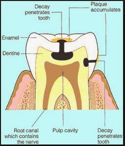
to support the process of remineralization. Tooth enamel daily undergoes acid attack that removes inorganic minerals from teeth. This is called demineralization. Remineralization is the opposite of demineralization. It occurs when inorganic molecules flow into a region of weakened enamel and make it stronger.

When the salivary flow is reduced, a chain of events occurs. The natural cleansing action is diminished, as are the buffering action and remineralization properties of saliva. People with diminished salivary flow experience a very fast rate of decay, many times faster and over several teeth. This type of dental decay is typically noted along the gumline, around existing dental work, and on exposed root surfaces.

Prevention
You can help prevent dental decay that can result from xerostomia:
  • Brushing and flossing correctly twice a day becomes very important.
  • Frequent sips of water during the day can help moisten the mouth and can help clear debris.
  • Daily use of a mouthrinse containing fluoride can help remineralize teeth.
  • Use a toothpaste containing sodium fluoride.
  • We recommend a daily brushing with a prescription, high-concentration sodium fluoride gel or paste. We will either dispense this or give you a prescription for it.
  • Chew sugarless gum or a rubber band to stimulate salivary flow.
  •  In moderate to severe cases, special fluoride delivery trays can be made for you to use at home. These will keep the high-concentration fluoride in a position to “soak” your teeth with fluoride for several minutes at a time.
  • We recommend that you have your teeth cleaned, polished, and an office-applied topical fluoride treatment every 3 months while the condition persists.

Dry mouth can have serious dental consequences and must be treated accordingly.
If you have any questions about xerostomia, please feel free to contact us at any of our three office locations:
Hymeadow: (512) 250-5012
Jollyville: (512) 346-8424
West William Cannon: (512) 445-5721

Monday, January 30, 2017

Prevention of Decay

Dental Decay
Dental caries (decay) is a bacterial infection, first of the enamel, then of the dentin of the tooth. The tradition in dentistry has been to surgically remove the diseased portion of the tooth by “drilling” out the decay and then filling the resulting hole in the tooth with some inert material. As most adults know, this procedure will be performed over and over again when new decay begins or when the filling (often silver) breaks or the tooth fractures.

Would it not be better to eliminate the cause of the infection and thus not be forced to have big holes drilled in the teeth? We believe the bacterial cause of the infection should be addressed.

Preventing the Risk of Dental Decay
There are several positive steps that you can take to reduce your risk of dental decay. First, all of the active decay in your mouth should be treated immediately. Next, all the teeth that would benefit from sealants should be treated. This will prevent bacteria from reaching into the pits, fissures, and grooves that normally exist on the occlusal (biting) surface of teeth. Any stray bacteria that may still be in the sealed area are effectively cut off from their food source and become inactive. Although sealants are most effective on teeth that have not been previously restored, they can be successfully placed on teeth filled with bonded fillings.

The infection can be treated with antimicrobials. We believe that the use of a fluoridated mouthrise twice daily or use of a prescription fluoridated dentifrice as directed provides great advantage. Not only is fluoride effective against bacteria but it also creates an environment that promotes remineralization of slightly damaged enamel. The decay process is reversed and the tooth may not have to be drilled. We may also prescribe a chlorhexidine mouthrinse, an antimicrobial oral rinse that has a great effect on Streptococcus mutans.

Your diet and oral self-care are important in dental decay prevention. When you eat junk food and drink sugary liquids, your teeth are more prone to decay. The more frequently you snack, the more prone your teeth will be to decay. If your brushing and flossing are not effective, your teeth will be more prone to decay. When you can’t brush after a meal, at least rinse your mouth with water within 15 minutes to dilute the acids forming from the ingested food or drink. If you have a diminished salivary flow, take frequent sips of water during the day to help dilute the acids produced by the bacteria.

If you have a continuing problem with active decay, we recommend more frequent preventative recare appointments. It has been repeatedly shown that patients who have good oral self-care and maintain a recare interval of 3 to 4 months have many fewer dentally related (cavities or gum disease) problems.

The routine 6-month recare interval is no longer our recommended schedule. That interval was based on a 50-year-old philosophy that never had any scientific basis! Times have changed. Present dental practice is based on proven scientific information. You might need to have your teeth cleaned by the hygienist twice each year or you may need to be seen more frequently.

For certain individuals, we also suggest testing the oral bacterial levels to determine the magnitude and presence of a Streptococcus mutans infection and to determine your risk level for future dental disease.

Wednesday, January 25, 2017

Implants, Crowns, and Bridges vs. Natural Teeth

Nothing can replace the natural teeth you were born with for chewing and function. However, very few people go through life without having teeth filled, crowns (caps) placed, or bridges and implants used to replace missing teeth. Crowns, bridges, and implants are the best answer and closest to your natural teeth, but they are not the same as healthy, natural teeth.

Crowns

Crowns are used to reconstruct a single tooth broken down by dental decay. Crowns are made of ceramic, resin, porcelain, porcelain plus metal, or resin plus metal materials. They are bonded or cemented onto the prepared tooth and cannot easily be removed from the tooth once placed. If the tooth was in good alignment before the crown was prepared, the crown will be in good alignment. If the tooth was misaligned before the crown, sometimes the crown may be made to obtain a more ideal shape and position. It is cleaned and flossed just like a natural tooth and is most like real teeth.

Bridges

Bridges are crowns that are attached together, suspending the crown portion of a false tooth in or over the space left by the missing tooth. A bridge can be used to replace one or several teeth. Sometimes a bridge is used to splint loose teeth together in order to make the teeth more stable. Bridges are usually made of metal covered with either porcelain or resin. Some of the newer bridges are made of all resin or all ceramic materials. They are cemented or bonded onto the existing prepared teeth and are not easily removed once placed. The bridge teeth can be brushed the same as natural teeth, but since they are attached together, must be flossed differently by using a floss threader or other device.
The teeth are generally the same shape as natural teeth. However, if the existing teeth (abutments) that are used to anchor the bridge have moved from their original position because a tooth or teeth have been missing for years, the added tooth (pontic) may be longer or shorter than the tooth that it is replacing. With a bridge, the false tooth will most often butt up against the soft tissue ridge where the removed tooth was.
The shape of the tongue side of the false tooth varies. It is usually smaller on the tongue side and completely fills the space. Food will have more of a tendency to collect in this area, so you must be prepared to clean it. If the missing tooth has been gone a long time, the ridge may have shrunk considerably, and the pontic tooth will be longer that the teeth on either side. If this is the case, there are several periodontal procedures that can be done prior to the construction of the bridge. These procedures will build up the tissue to its former height. The more your mouth has changed from its normal state, the harder it is to make new teeth look and feel natural.

Implant Crowns

Implant crowns are used to replace single or multiple missing teeth. They are either cemented or screwed-retained onto an implant fixture. The crowns are made of porcelain or resin and metal. But they have some significant differences from the natural teeth they replace. Teeth are supported by a root or roots that are irregular in shape. Implants are round. The cross-sectional of the implant will never match that of the tooth it is replacing. A multirooted tooth may be replaced by a single implant, so the manner in which an implant crown comes out of the soft tissue ridge will appear different from a natural tooth. There will be more space between the implant root and the adjacent teeth. Implant crowns are often cemented with temporary cement. This allows the dentist to easily take off the crown and evaluate how the implant is doing. Crowns on teeth are usually cemented with a final cement. Implant-supported crowns are wonderful, but not the same as natural teeth with crowns. Be prepared for some differences. Expect more maintenance on your part and in the dental office with crowns, bridges, and implants.


Smokers take note: There is a heightened risk of dental implant failures among smokers—as much as 20% greater failure rate!

Monday, August 4, 2014

Dentin Decay

Detect decay by inspecting the color and hardness
Usually, dental decay is fairly easy to detect. When a cavity is just beginning, it is typically identified by a brown or white color or a change in the translucency of the enamel of the tooth. The dentist or dental hygienist uses a special dental instrument called an explorer to feel the suspect area and check its hardness. If the area is hard, in other words, if no break in the enamel layer is detected, we feel there is not a cavity present. If, however, the surface feels soft and the explorer "sticks" in the suspect site, we feel a cavity is present.

Fluoride makes our enamel stronger, but dentin decay is harder to detect Because of the widespread use and availability of fluoride in our drinking water, foods, and oral care products, we are seeing a different decay pattern. The appearance is different from the typical pattern of decay and more difficult to detect. As the outer surface of the enamel absorbs fluoride (from toothpaste, for example), the enamel becomes very resistant to demineralization and eventual decay. If there is a small break in the integrity of the enamel, a pit or groove where decay-causing bacteria can live, the bacteria can dissolve the enamel in such a way that the hole in the enamel cannot be detected. Once the decay-causing bacteria reach the underlying dentin, the acids eat away at that substance and quickly make a large cavity - but one that still cannot be easily seen or detected. In this way the enamel becomes undermined. A dentist looking at such a small cavity would think it very easy to restore. However, once the decayed portion of the enamel is removed and the dentin becomes visible, the true extent of the damage becomes obvious. The small cavity becomes a big cavity.

One of the major problems with decay that appears to occur only in the dentin is in the detection. If a radiograph is taken a part of the periodic examination process, we may be able to see dentin decay if it is moderate to extensive. Decay seen on radiographs is typically two to seven times greater in the tooth. Modern high-speed radiographic film and reduced x-ray exposure makes it more difficult to detect early decay on radiographs.

You can prevent decay by using fluoride
The conscientious application of a source of topical fluoride, through either an over-the-counter dentifrice or a prescription fluoride product, and thorough plaque removal are essential. Bonded sealants are also an effective protection against dentin decay. We strongly advice these procedures. Periodic examinations at intervals recommended by the dentist catch decay at the earliest possible time. This is the only way to keep small problems from developing into larger problems.

Click here to check out our blog post on foods that cause decay. 
Click here for our post on how to prevent decay.

If you have any questions about dentin decay, please feel free to ask us. 

Wednesday, July 30, 2014

Sealants and Fluoride: Benefit to Adult Patients

Dental decay can develop at any time, regardless of a person's age. A change in diet, change in lifestyle, change in oral self-care habits, the use of prescription medications, or a change in systemic health due to the normal aging process can all affect the caries (decay) susceptibility. Few people remain completely free of decay. Proper oral self-care on your part and properly spaced dental hygiene prevention appointments will go a long way to reduce the opportunity to have new decay to begin.

As you age, it is possible that some of your gum tissue will recede, exposing the root surfaces of your teeth. This gum recession can occur from improper brushing (brushing too hard with a hard toothbrush) or as a result of past periodontal problems. The more a tooth and root are exposed, the greater is the surface area you will have to keep clean. Sometimes the teeth with exposed roots are very hard to keep clean. These roots may be sensitive to temperature changes and are often times uncomfortable to brush. Decreased salivary flow (dry mouth) helps to create a breeding ground for bacteria to accumulate on the enamel and especially on the root surface. And root decay usually progresses quite quickly!

Goal of Prevention
Your goal should be to keep the dentist from drilling your teeth. Any reasonable preventive measure that is available should be seriously considered. When the dentist drills, you lose. When the dentist does not drill, you win.
                               
                                                         Dental Sealants
Please see our blog post on sealants. Although sealants are primarily designed for children, adults who have a history of active decay should consider having sealants placed on the posterior (back) teeth where indicated. We will tell you where it is possible to place the sealants. Even if you have not had a cavity for a long time, consider the application of a sealant as an inexpensive insurance policy for your teeth. Perhaps you would never get decay on the unsealed surfaces. But, just as you insure your home against destruction by fire, a sealant insures the tooth surface from decay. Preventive measures may allow you to avoid having your teeth drilled. You win! 

Topical Fluoride
For a similar reason, we advise the use of topical fluoride treatments for adults. The effectiveness of systemic and topical fluoride in preventing decay is well documented. When a cavity first starts, an application of fluoride might (depending on when it is used) reduce or eliminate the need for drilling.
Fluoride Varnish

An alternative to the fluoride treatment we can provide in our office is a daily rinse. If you can rinse with an over-the-counter mouth rinse containing fluoride every night as directed on the rinse label, you do not need the office topical fluoride treatment. If you cannot rinse daily as instructed, you will need the benefit from the strong office-applied topical fluoride treatment. Your oral health will benefit most from small increments of fluoride that are applied daily rather than one larger concentration every 6 months. However, only you know whether you will be faithful in your rinsing routine. When in doubt, let us do it here.

We stress prevention of dental disease above all else. Sealants and topical fluoride treatments are two of the more important preventive dental measures that we believe will significantly enhance your oral health.

Tuesday, July 29, 2014

Seal the Deal: Everything You Need To Know About Sealants

What are dental sealants?  

Dental sealants represent one of the greatest advances in modern dentistry. Unfortunately, too many children do not receive the protective benefits of dental sealants. Dental sealants are clear protective coatings that are easily applied; they cover the tooth surface, preventing bacteria and food particles from settling into the pits and fissures (grooves) of the teeth. It is a thin coat of plastic that is painted on the surface of teeth, usually the premolars and molars in order to prevent caries.

Decay on back teeth, premolars, and molars usually begins in the grooves and fissures that normally exist on the biting surfaces of the back teeth.  Dental sealants, available since the 1960s, are clear plastic coatings that can be placed on the biting and grinding surfaces of posterior teeth.  These sealants prevent the formation of decay on the treated surfaces.  Sealants can even be placed of teeth with small areas of decay known as incipient carious lesions.  The sealants will stop the customary progress of tooth destruction.  

Effectiveness
It can remain on the tooth from 3 to more than 20 years, depending on the tooth, type of sealant used, and the eating habits of the patient.  It can only be placed on teeth that have not been previously restored.
It is well known that the use of fluoride increases the resistance of enamel to decay. Unfortunately, the pits and fissures of the teeth do not benefit from the effects of fluoride as greatly as smooth enamel surfaces do. Scientific studies have proved that properly placed dental sealants are 100% effective in protecting development of cavities in sealed tooth surfaces. 

Why sealants are necessary
The narrow width and uneven depth of pits and fissures make them ideal places for the accumulation of food and acid-producing bacteria. Saliva, which helps to remove food particles from other areas of the mouth, cannot clean deep pits and fissures. Pits and fissures on the teeth are so tiny that even a single toothbrush bristle is too large to enter for cleaning purposes. Another difficulty associated with deep pits and fissures is that the enamel that lies at the base of the fissures is thinner than the enamel around the rest of the tooth. This means that not only can deep narrow fissures make it more likely that tooth decay will occur, but any decay that does form will penetrate through the thin enamel and progress more quickly into the pulp.

We, at this office, are dedicated to the prevention of oral disease.  It is clear that if the initial decay is prevented from beginning or is small enough to use a sealant, there is a great savings in time, money, discomfort, and tooth structure.  Decayed teeth must have the decay removed by drilling, then they must be filled.  This drill and fill may have to be done several times over the patient's lifetime as the filling ages and needs replacement.  We strongly suggest that patients who have teeth that can be successfully protected with a sealant material consider having this procedure performed as soon as possible.


How sealants work
The sealant acts as a physical barrier that prevents decay. Small food particles and plaque (bacteria) cannot penetrate through or around a sealant. As long as the sealant remains intact, the tooth is protected. However, if part of the sealant or bond is broken, the sealant protection is lost. 

A study completed in 1991 found that one application of sealant reduced biting surface decay 52% over a 15-year period.  Another study, completed in 1990, showed that decay on biting surfaces could be reduced 95% over 10 years if 2% to 4% of the sealants were routinely repaired each year.  We expect sealants to last many years. After a sealant is applied it can last up to ten years, however regular check-ups are recommended since they can chip or wear off. Replacing or repairing sealants, as needed, on an ongoing basis will give the best protection. 

A sealant is not meant as a substitute for proper brushing and flossing habits. The effectiveness of the sealant is reduced if oral self-care is neglected. Also, cavities can still form on untreated surfaces. Therefore, a topical fluoride treatment remains an essential and necessary preventative aid.

Application of sealants
The sealant is placed on the tooth through a chemical/mechanical bonding procedure.  There is not drilling or local anesthesia required for the sealant application procedure.  It is entirely painless.

Problems with sealants
On occasion, teeth with very small initial carious lesions may be inadvertently sealed, or some bacteria may remain beneath the sealant. It was one believed that if this were to occur, decay would develop under the sealant. However, numerous studies have shown that this does not occur. Bacteria cannot survive beneath a properly placed sealant because the carbohydrates that they need to survive cannot reach them. Studies have shown that the number of bacteria in small, existing carious lesions that had been sealed actually decreased dramatically over time. The most important factor is that the sealant must be properly placed.

When sealants are used
Sealants are used primarily on children, but in certain circumstances, adults also can benefit from their use. Children and teenagers are the best candidates for sealants. Children are prone to caries from age 6-14 and applying a sealant as soon as a child’s permanent molars and premolars erupt can be a great way to protect your child from tooth decay. Some maxillary central and lateral incisors may have deep pits that could be protected by sealants. Sealants are indicated for teeth with deep pits and fissures, preferably in recently erupted teeth (i.e., within previous 4 years). Sealants should be used as part of a prevention program that includes the use of fluorides, dietary considerations, plaque control, and regular dental examinations.

We especially advise that children have the sealant applied to their teeth as soon as the teeth break through the gum and the biting surfaces of the teeth are no longer covered with gum tissue.  If the teeth cannot be totally isolated from the moisture in the mouth during the bonding process, it is likely that the sealant will not remain on the tooth for as long a period of time as expected.  The sealant is most often applied to permanent teeth, but sometimes a situation arises in which it would be beneficial to have the sealant applied to a primary tooth.

In both 1984 and 1994, sealants have been recommended by the U.S. Public Health Services and Surgeon General of the United States, among others. We know that sealants are one of the most important treatments available for prevention of dental decay.

If you have any questions about sealants, please feel free to ask us.

Thursday, July 24, 2014

Sensitive Teeth

Teeth can become sensitive for many reasons. Sometimes, the sensitivity is an indication of a potentially serious problem. Other times, the dentally related problem may be small but the effects (the sensitivity) are extremely aggravating. 

What do we mean when we say "sensitive teeth?"
Twinges of pain or discomfort in our teeth in certain situations such as drinking or eating cold things, drinking or eating hot things, eating sweets, and touching the teeth with other teeth or the tongue. Your symptoms will involve reactions to temperature or pressure. Sensitivity to cold drinks or foods is the most common symptom. 

Two types of sensitivity:

1. Dentinal sensitivity
When the dentin (middle layer) of a tooth is exposed. Normally, dentin is covered by enamel. Dentin becomes exposed when the outer protective layers of enamel wear away. Dentin contains tiny openings called tubules. Inside each tubule lies a nerve branch that comes from the tooth's pulp (the nerve center of the tooth). When the dentin is exposed, cold or hot temperature or pressure can affect these nerve branches, causing sensitivity.

Causes:
  • Inadvertent notching of the tooth surface and/or recession of the gum tissue (exposing the root surface of the tooth) caused by improper brushing: either brushing too hard, brushing with a toothbrush that is too hard, or using an improper brushing technique, which will wear away the enamel layer. This sensitivity can range from mild to extreme; the degree of sensitivity does not appear to be related to the size of the root exposure or notch.
  • Poor oral hygiene, allowing tartar to build up at the gum line
  • Untreated cavities
  • An old filling with a crack or leak
  • Receding gums that expose the tooth's roots, often caused by periodontal disease
  • Gum surgery that exposes the tooth's roots
  • Tooth whitening in people who have tooth roots that already are exposed
  • Frequently eating acidic foods or drinking acidic liquids 
  • Purposeful repositioning of the gum tissue during gum surgery can also lead to tooth sensitivity. While recession from brushing is slow, gum recession following gum repositioning occurs very quickly. The portion of the tooth once covered with gum and bone may now be exposed.  Root sensitivity in these instances can be quite severe and immediate. It can sometimes last four months or years if not treated.
2. Pulpal sensitivity
The reaction of the tooth's pulp. The pulp is a mass of blood vessels and nerves in the center of each tooth. Pulpal sensitivity affects only a single tooth.
Causes:
  • Decay or infection. When a tooth is decayed, temperature changes and sweets will make it sensitive. The solution can be as simple as removing the decay and placing an appropriate restoration.
  • A recent filling. A tooth may be sensitive to cold for several weeks after you get a filling. The metals in amalgam (silver) conduct the cold very well, transmitting it to the pulp. Bonded (tooth-colored) fillings require etching the tooth with acid before the filling is placed. In some cases, this etching removes enough enamel to make the tooth sensitive. If a filling is defective or failing, leakage around the filling may cause the tooth to become sensitive. The solution can be as simple as removing the defective filling and placing an appropriate restoration.
  • Excessive pressure from clenching or grinding
  • A cracked or broken tooth. If the tooth is fractured, you may be sensitive to temperature changes or when chewing food. This fracture condition may be hard to diagnose. If you think you might have this type of sensitivity, read our blog post on "cracked tooth syndrome."
  • A tooth can become sensitive after it has been prepared (drilled) for a restoration (filling). You may have been anesthetized during the procedure, so you did not feel any discomfort when the nerve in the tooth reacted to the heat generated by the drill. The closer the drill comes to the nerve, the more likely it is to cause a sensitivity problem. The high-speed rotation of the bur in the drill generates heat, and the response of the nerve to heat is inflammation. This inflammation is felt by you as a "sensitivity."  If the decay, fracture, or drilling was not too deep, this sensitivity will decrease over time. A week to a month or two is not an unusual length of time for the sensitivity to disappear. A good sign is the continued decrease of sensitivity.  However, if the occlusion (bite) is off after the restoration has been placed, the tooth may either become sensitive or may stay sensitive. Once the bite is adjusted, though, the sensitivity should disappear.
  • A dying nerve. This can be the result of a deep cavity. Commonly, the sensitive tooth holds an old large filling. The nerve may be damaged during drilling and the nerve has been dying gradually ever since. If this is the problem, the tooth will need endodontic treatment (root canal treatment).
See this post about the 10 biggest causes of tooth sensitivity.

How will the doctor diagnose sensitivity?
Your dentist will look at your dental history and will examine your mouth. You also will need x-rays to show if there is decay or a problem with the nerve. The dentist will ask about your oral habits. Grinding or clenching your teeth can contribute to sensitivity. Your dentist also will look for decay, deep fillings, and exposed root surfaces. During the consultation, your dentist uses an instrument called a spray gun to dispense air across every area of each tooth in order to locate the sensitivity. He or she may use an explorer - a metal instrument with a sharp point - to test teeth for sensitivity.
 
How will it be treated?
  • Your dentist or dental hygienist will clean your teeth. If your teeth are too sensitive to be cleaned, your dentist may use a local anesthetic or nitrous oxide before the cleaning. 
  • If there is a notch in the tooth or the shape of the defect is appropriate, the defect is restored (filled in) with a bonded material. This can give immediate relief -- sometimes partially, sometimes fully. 
  • When there is no defect to be restored, the exposed and sensitive root surfaces are covered with a dentin-bonding or other material. This material is invisible and has very little thickness, so you do not notice any change in the appearance of the tooth; but it works. It may have to be reapplied after several months because the bonding material has worn away by tooth-brushing.
  • If you have pulpal sensitivity and the tooth's nerve is damaged or dying, you will need a root canal. Your dentist will remove the nerve and place a non-reactive substance in the space where the nerve was. The tooth will no longer have a continuous barrier of enamel to protect it. Therefore, it will be restored with either a composite filling or a crown.
  • Your dentist may apply a fluoride varnish to protect your teeth. This temporarily reduces sensitivity and strengthens your teeth.
  • Using fluoride toothpastes and fluoride mouth rinses are home will help to reduce sensitivity. You can also buy toothpastes just for sensitive teeth. You should choose a fluoride mouth rinse that uses neutral sodium fluoride.
How can I prevent it?
If you have any questions about tooth sensitivity, please feel free to call our office for a consultation at (512) 250 - 5012.

Tuesday, July 15, 2014

Prevention of Decay

What is dental decay, and how do dentists fix it?
Dental caries (decay) is a bacterial infection, first of the enamel, then of the dentin of the tooth. The tradition in dentistry has been to surgically remove the diseased portion of the tooth by "drilling" out the decay and then filling the resulting hole in the tooth with some inert material. As most adults know, this procedure will be performed over and over again when new decay begins or when the filling (often silver) breaks or the tooth fractures.

Click here to learn about decay in young children.

Why should I practice prevention?
Would it not be better to eliminate the cause of the infection and thus not be forced to have big holes drilled in the teeth? We believe the bacterial cause of the infection should be addressed.

How do I prevent dental decay?
There are several positive steps that you can take to reduce your risk of dental decay. 

A. At the dentist's office
1. First, all the active decay in your mouth should be treated immediately. 

2. Next, all teeth that would benefit from sealants should be treated. 
  • Why? This will prevent bacteria from reaching into the pits, fissures, and grooves that normally exist on the occlusal (biting) surfaces of teeth. Any stray bacteria that may still be in the sealed area are effectively cut off from their source of food and become inactive. Although sealants are most effective on teeth that have not been previously restored, they can be successfully placed on teeth filled with bonded fillings.
3. The infection can be treated with antimicrobials. 

4. We believe that the use of a fluoridated mouthrinse twice daily or use of a prescription fluoridated dentifrice as directed provides a great advantage. 
5. We may also prescribe a chlorhexidine mouthrinse, an antimicrobial oral rinse that has a great effect on Streptococcus mutans.

B. In your daily life
6. Your diet and oral self-care are important in dental decay prevention. When you eat junk food and drink sugary liquids, your teeth are more prone to decay. The more frequently you snack, the more prone your teeth will be to decay. 

7. If your brushing and flossing are not effective, your teeth will be more prone to decay. When you can't brush after a meal, at least rinse your mouth with water within 15 minutes to dilute the acids forming from the ingested food or drink. 

8. If you have a diminished salivary flow, take frequent sips of water during the day to help dilute the acids produced by the bacteria.

What if decay keeps coming back?
If you have a continuing problem with active decay, we recommend more frequent preventive recare appointments. It has been repeatedly shown that patients who have good oral self-care and maintain a recare interval of 3 to 4 months have many fewer dentally related (cavities or gum disease) problems.

Present-day dental suggestions are based on proven scientific information. You might need to have your teeth cleaned by the hygienist twice each year, or you may need to be seen more frequently. 

For certain individuals, we also suggest testing the oral bacterial levels to determine the magnitude and presence of a Streptococcus mutans infection and to determine your risk level for future dental disease.

If you have any questions about the prevention of dental decay, please feel free to ask us.

Monday, July 14, 2014

Extractions

Reasons for Recommending Tooth Extraction
Teeth may need to be extracted for several reasons, including but not limited to:
  • severe periodontal disease
  • irreversible damage to the nerve tissue inside the tooth (and the patient decides against saving the tooth)
  • failed endodontic therapy
  • extreme fracture or decay of the tooth structure
  • improper positioning of the tooth or for orthodontic purposes
To a great extent, the reason for the extraction will influence the amount of discomfort you might experience subsequent to the procedure. When the tooth is to be extracted for periodontal reasons, there will be reduced bone support for the tooth and the tooth might be removed more easily than if there were full bone support. In this case, there might be lessened discomfort following the extraction.

The Procedure
1. Numb the tooth: Your dentist will need to anesthetize (numb up) both your tooth and the bone and gum tissue that surround it. This is done as an injection.

2. Extraction: The root portion of a tooth is firmly encased in bone (its socket), and tightly held in place by a ligament. During the extraction process, the dentist needs to both "expand the socket" (widen and enlarge it) and separate the tooth from its ligament, to the point where the tooth is loose and free to come out.
  • What does it mean to "expand" a tooth's socket? If you have ever tried to remove a tent stake that has been driven deeply into the ground, you know that you can't just pull the stake straight up. Instead, you first have to rock the stake back and forth, widening the hole in which it is lodged. Once the hole has been enlarged enough, the stake will come out easily.
  • How does the dentist expand the jaw bone? The bone inside the jaw is spongy. When a dentist applies firm pressure to a tooth (forcing it against the side of its socket), the bone will compress. After repeated application of pressure, from many different angles, the entire socket gradually becomes expanded. At some point, enough space will have been created (and the ligament separated from the tooth enough) that the tooth will come out.
3. Closing the extraction site: Once your tooth has been removed, your dentist will begin the process of closing your extraction site. This may include:
  • Removing infected or pathologic tissue by curetting (scraping) the walls of the tooth socket
  • Using finger pressure to re-compress the "expanded" socket
  • Rounding off sharp bone edges
  • Evaluating the tooth socket for sinus complications (upper back teeth)
  • Washing out ("irrigating") the socket, so to remove any loose bone or tooth fragments that remain
  • Placing stitches (usually only after surgical extractions).
  • Placing folded gauze over your extraction site and then having you bite down on it so to create firm pressure
The Tools
1. Dental Elevators: During the extraction process, a dentist will usually use an elevator first. These instruments are designed to be wedged in the ligament space between the tooth and its surrounding bone. As the elevator is forced and twisted, the tooth is pressed and rocked against the bone. This helps to expand the socket. It also helps to separate the tooth from its ligament. As this work is continued, the tooth will become more and more mobile. In some cases, the elevator may be able to shove the tooth on out. If not, the dentist will switch to the use of extraction forceps and remove the tooth with them.

2. Extraction Forceps: A dentist will usually keep a number of different extraction forceps on hand. Each one will have a design that's been specially made to grasp a certain type of tooth. When they're used, the dentist will grasp the tooth with the forceps and them firmly and deliberately rock it back and forth as much as it will. Because the bone that surrounds the tooth is compressible, the socket will expand. In addition to a rocking motion, a dentist will also rotate the tooth back and forth. This twisting action helps to rip and tear the tooth from the ligament that binds it in place. At some point, the socket will be enlarged enough, and the ligament torn enough, that the tooth can be easily removed.

What will I feel?
1. Pressure
2. You should not feel any pain 
3. Expect to hear startling extraction noises: These are often just routine

Following the Extraction
We will tell you the reason for the extraction and let you know what to expect following the procedure. Please follow the instructions given to you. If antibiotics are prescribed, take them until the prescription is completely finished. If pain medication is prescribed, take it only if necessary. If the medication prescribed contains a narcotic component, such as codeine, do not drive a motor vehicle or operate machinery that could prove dangerous to yourself or others. Expect some bleeding to occur from the extraction site for the first 24 hours. Remember, there is now a hole in your jaw from which the tooth has been removed, and the hole can be quite large. Some bleeding is to be expected.
  • Do not spit, rinse, or smoke for 24 hours
  • Do not drink through a straw for 24 hours
  • It would be a good idea not to brush near the extraction site for a day or two
  • When you brush and floss the area, be gentle!
  • For 24 hours after the extraction, try to chew food away from the extraction site
  • Some slight swelling in the area is to be expected, especially if the extraction was difficult
  • If sutures are placed, return to have them removed
  • If medication has been prescribed for possible post extraction discomfort, take it as directed
  • If prescription medication has NOT been given, you may take your usual over-the-counter pain reliever, as directed
 Please notify us if:
  • There is extended bleeding from the extraction site. Slight bleeding for several hours is normal.
  • Anything other than slight swelling occurs.
  • Discomfort continues for more than 24 hours, especially if it is not relieved by over-the-counter pain relievers.
Some infrequent complications of routine oral surgical procedures include (but are not limited to):
  • fracture of adjacent teeth or restorations (which of course would mean that these affected areas must be restored to normal function after the healing of the extraction site)
  • separated root tips or root fragments
  • temporary or permanent nerve damage to the area, resulting in anesthesia or paresthesia (numbness)
  • incomplete healing, resulting in severe pain - a "dry socket"
  • fracture of the surrounding bone
If you have any questions about reasons for dental extraction, please feel free to ask us.

Friday, July 11, 2014

Gingival Grafts

What is it?
Periodontal (gum) surgery is most often associated with the removal of soft tissue. But there are times when it is necessary to use soft tissue to cover an area that has too little soft tissue remaining. 

What causes it?
Exposed roots may be due to the improper brushing, periodontal disease, or genetic structure. Brushing too hard and/or with a hard toothbrush can cause the gingival tissue to disappear. The gingival margin (gumline) changes, and one or more millimeters of root structure can be exposed. Active periodontal disease can cause loss of this soft tissue too. 

Why you should care
This exposure can lead to teeth that are very sensitive to temperature changes, root decay, or are quite unsightly. The disfigured soft tissue line can lead to a plaque trap, causing more disease and further problems.


How dentists fix it
Two methods of resolving these problems are free gingival grafts and attached gingival grafts. Both of these are periodontal surgical procedures. In both cases, a local anesthetic will be used to numb the treatment site. 
In the case of an attached graft, gingival tissue is taken from the donor site and moved to the area where root coverage is required. The graft tissue is sutured into place, and a dressing is placed over the treated area. The dressing remains in place for several weeks and is then removed. An attached graft is not completely removed from the donor site. The donor site is adjacent to the site that needs root coverage. There is an incision made in the gum tissue, and the tissue is moved sideways, up, or down and sutured into place. Again, a protected dressing is placed to protect the area while healing occurs. Any anticipated postoperative discomfort is resolved with medication. Most discomfort will come from a free graft donor site.

Who performs the procedure?
A periodontist (gum specialist) usually performs these procedures, as well as other types of grafts and root coverage treatments. After the periodontist examines the areas needing treatment, you will have a better idea of what treatment will consist of, what the appearance may be, anticipated healing times, postoperative discomfort, and cost.

How can you prevent it?
Most patients have heard that they should take better care of their teeth - brush and floss the teeth. Some patients brush too hard and in a back-and-forth motion; and some facial, bony structures and biting forces are such that root exposure happens, resulting in longer looking teeth. The grafting procedures mentioned here can restore the proper gingival marginal architecture, prevent root decay, reduce or eliminate thermal sensitivity, and make your smile look great again.

If you have any questions about gingival grafts, please feel free to ask us.

Wednesday, July 9, 2014

Foods That Cause Tooth Decay

Cariogenic: causing tooth decay.
Foods (such as candy and sweets) that contain sugar and other carbohydrates that can be metabolized by bacteria in plaque are described as cariogenic

How long food remains in mouth:
  • Sugary liquids, such as soft drinks, leave the mouth quickly and are not as cariogenic
  • Sticky foods, such as raisins or caramels, adhere to the teeth and stay in the mouth longer and are more cariogenic
Frequency vs. Amount:
  • Someone who snacks all day on small amounts of cariogenic food is far more likely to develop decay
  • Someone who eats a larger amount of cariogenic food, but only once a day, is less likely to develop decay 
Refined carbohydrates vs. Complex carbohydrates:
  • Foods such as crackers, although not sweet, are cariogenic because they contain refined carbohydrates that stick to the teeth. They remain in the mouth long enough to be broken down into sugars that can be used by the bacteria in plaque.
  • Complex carbohydrates, such as fruits and vegetables, are less cariogenic because they clear the mouth before they are converted into simple sugars that can be used by bacteria.  
Flow of saliva to speed clearance of food from mouth and provide a source of dietary fluoride to strengthen the tooth and assist in remineralization
  • Sugarless gum or hard candy, as well as sipping water frequently, will increase the saliva flow
  • Various medicines can cause a dry mouth, and try to avoid spicy or salty foods

Tuesday, July 8, 2014

Give Yourself A 60-Second Dental Check-Up

Regular visits to your dentists are absolutely essential to prevent serious dental problems. But it is also important to give yourself an oral check-up from time to time. This simple process just takes a minute, but it can yield valuable clues to the health of your mouth, teeth, and gums.

Stand before a well-lit mirror and open your mouth wide.
1. Check your gum line and the lines between your teeth
Good: There should be no brown lines here
Bad: If you see brown lines, that is a sign of tartar build-up
What to do: Time to go to the dentist. Once the brown lines are cleaned up at the clinic, regular flossing will keep tartar away.

2. Stick out your tongue. What does the surface look like? 
Good: A healthy tongue should be pink
Bad: If it is coated with white, it can be an indication of poor digestion. This coating also results in bad breath.
What to do: Use a stainless steel scraper to clean your tongue every morning. 

3. Are there ridges along your tongue? 
Bad: This is often a sign of tooth grinding. People may not be aware they grind their teeth, but it can happen when they are stressed. 
What to do: If you wake up with an aching jaw, you should see a dentist.

4. Take a look at the inside of your cheeks. 
Bad: Do you see white patches? These can occur if you chew the inside of your mouth when you bite and chew your food. 
What to do: These should be checked by your dentist because a wound inside the cheek can not only get infected, but may also be a sign of pre-cancerous changes, especially in smokers. An ulcer that does not heal within three weeks should also be investigated.

5. How do your gums look?
Good: If they are pink and rosy, your gums are in good health. 
Bad: But if they are red, it is a signal that gingivitis or gum inflammation has turned into gum disease. Another clue: gums that bleed when you brush your teeth. 
What to do: Your dentist can help you with a course of antibiotics and dental hygiene guidance on how to rebuild gum health. 

Bad: Pale gums can indicate iron deficiency - a warning sign of anemia. 
What to do: Up your intake of green vegetables, tofu, chickpeas, lentils, and dried fruits such as raisins and prunes.